
The World at a Micro Level: Seeing What Mines Researchers See
Researchers across the Mines campus often see a part of the world that the average person will never know. Fortunately, using advanced microscopy, researchers were willing to share a peek at what they are working on, allowing us to see what they see while in the lab.
3-D transmission x-ray microscopy image of platinum nickel nanowire fuel cell catalyst when embedded in an electrode.
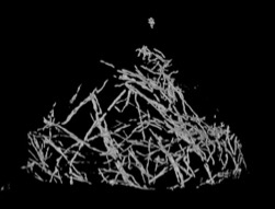
Nanoscale chemical mapping of a catalyst for liquefying natural gas.
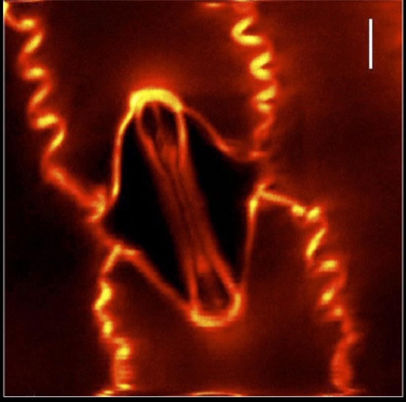
Two photon excitation fluorescence image of a Yellow Fluorescent Protein labeled corn plant.
Two photon excitation fluorescence image of a Yellow Fluorescent Protein labeled corn plant.
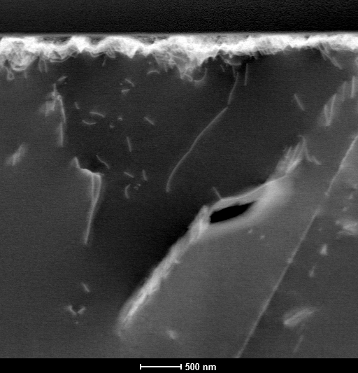
Scanning transmission electron microscopy micrograph illustrating defects in a natural Brookite (Titanium Dioxide) crystal acquired in dark field mode. Crystallographic defects include line defects such as dislocations and planar defects such as stacking faults and grain boundaries.
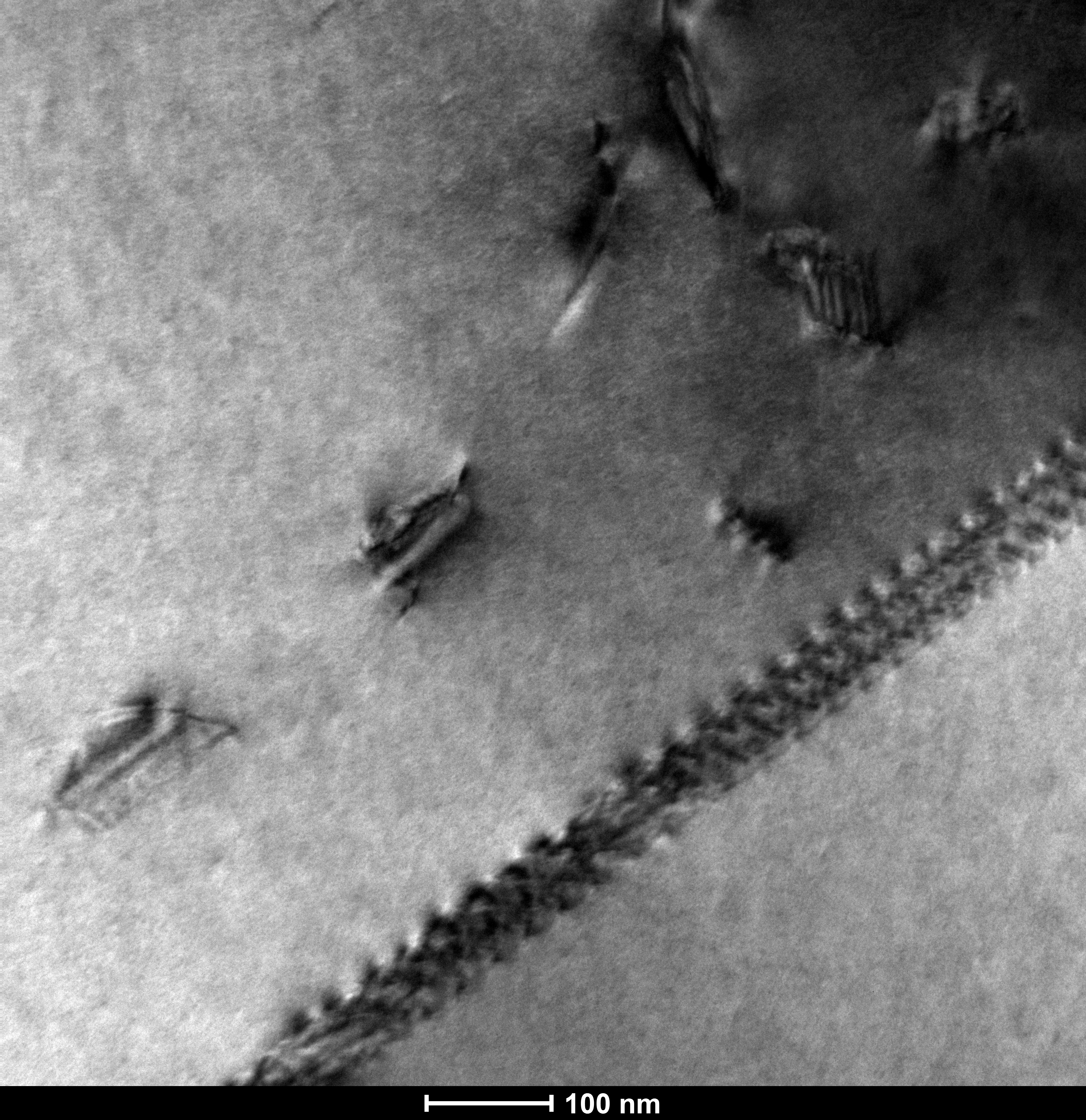
Transmission electron microscopy image illustrating defects in a natural Brookite (Titanium Dioxide) crystal acquired in dark field mode. At bottom right, a series of dislocation loops are present, giving the appearance of a zipper. Defects in the top left of the image are single dislocation loops.
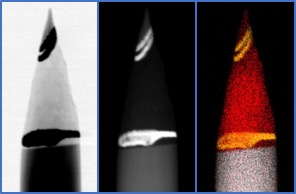
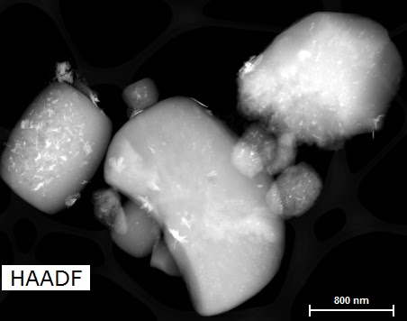
Thinned, needle-shaped sample for atom probe tomography, prepared by milling in a dual-beam scanning electron microscope and focusedion beam (FIB) instrument. Left to right: Scanning transmission electron microscopy images in bright-field and high-angle annular dark field modes, with a corresponding energy dispersive spectroscopy compositional map. The carbon sample (red) is clearly sandwiched between layers of metallic gold coating (yellow) on a silicon substrate (gray).
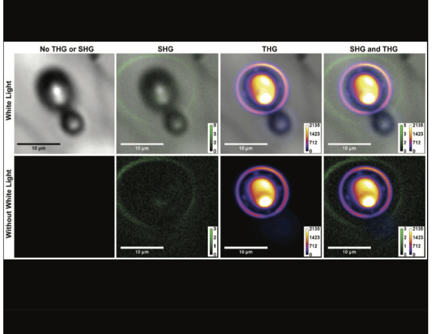
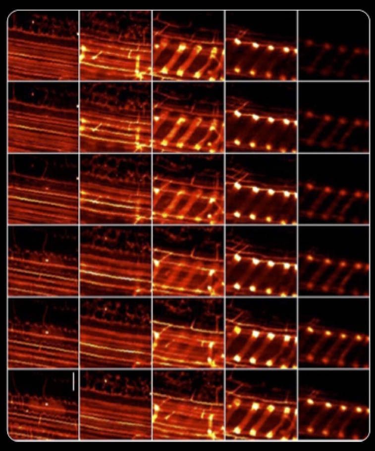
Image series of bubbles in glass that were created by a femtosecond laser. Two different image contrast mechanisms are used, second harmonic generation (SHG) and third harmonic generation (THG). These bring out different features—especially when compared to the traditional white light image on the far left column.


